Assessing the Influence of Microarchitecture on the Mechanical Performance of Bone and its Changes in Microgravity from in-vivo Measurements (Phase III)
2D and 3D Quantification of Bone Structure and its Changes in Microgravity Condition by Measures of Complexity (Phase I, II)
European Space Agency (ESA) Contract: 14592, MAP Project Number AO-99-030 / AO-2004-125, Project acronym: Bone3D
Have a look at our Gallery where you can find some movies.
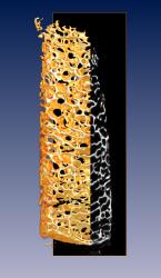
Visualization of a 3D data set obtained by micro-CT of a human tibia bone biopsy (Visualization program: Amira) | 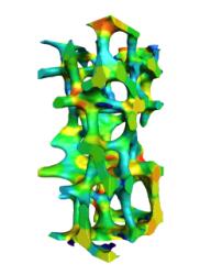
Color coded part from a tibia bone biopsy, visualized by Amira from a data-set acquired by micro-CT |
Table of Contents
- 1. Project Description
- 2. Participating Research Groups
- Max Planck Institute of Colloids and Interfaces, Department of Biomaterials, Golm, Germany
- Institute of Anatomy, University of Aarhus
- Group Nichtlineare Dynamik, University of Potsdam
- Scientific Visualization Department, Zuse Institute Berlin (ZIB)
- Center of Muscle and Bone Research, Free University Berlin
- Ludwig Boltzmann Institute of Osteology
- 3. Documents
- 4. List of Addresses
- 5. Gallery
| Coordinator: | Peter I. Saparin, PhD | Max Plack Institute of Colloids and Interfaces, Golm, Germany |
| Wolfgang Gowin, MD, PhD | Former Coordinator til July 31, 2004 | |
| Team Members: | Peter Fratzl, PhD | Max Plack Institute of Colloids and Interfaces, Golm, Germany |
| Richard Weinkamer, PhD | Max Plack Institute of Colloids and Interfaces, Golm, Germany | |
| Dieter Felsenberg, MD, PhD | Free University Berlin, Germany | |
| Gisela Beller, MD | Free University Berlin, Germany | |
| Jürgen Kurths, PhD | University Potsdam, Germany | |
| Norbert Marwan, PhD | University Potsdam, Germany | |
| Jesper Skovhus Thomsen, PhD | University of Aarhus, Denmark | |
| Hans-Christian Hege | Zuse Institute Berlin (ZIB), Germany | |
| Steffen Prohaska | Zuse Institute Berlin (ZIB), Germany | |
| Bruno Koller, PhD | Scanco Medical AG, Switzerland | |
| Thomas von der Haar, PhD | Siemens AG, Forchheim, Germany | |
| Christian Asbeck | Siemens AG, Forchheim, Germany | |
| Malte Westerhoff | Indeed - Visual Concepts, Berlin, Germany | |
| Project User Group: | Roche Pharmaceuticals, Switzerland | |
| Hewlett Packard (HP), Germany |
After the contract was signed on December 14, 2000; the project started at the beginning of January 2001. The first phase was successfully completed by the end of December 2002. These first two years are documented in the Final Report. The project team applied successfully for a three-year-extension, and received peer-reviewed approval for that in December 2002. The second phase started in January 2003 and will last until the end of September 2005.
The project develops the tools to evaluate structural loss in bone architecture and gains new quantitative information about the bone metabolism in microgravity condition. This evaluation is mostly based on symbolic dynamics and measures of complexity derived from the field of nonlinear dynamics. The most precise procedure for diagnosing structural alterations will be developed and scientifically proven through the research project. The outcome of such a comprehensive diagnostic program will provide the fundamental basis for monitoring, prevention, and treatment of structural changes of the bone in microgravity condition. This will have an impact on space-flying personnel and on patients with bone diseases on Earth.
The following tasks have been solved during the first phase of the project:
development of a method for non-invasive evaluation of the bone structure using 2D quantitative computed tomography images based on human specimens acquired at different skeletal sites,
development of a procedure for the precise quantification of a 3D bone architectural composition by analyzing human bone biopsy datasets obtained by micro-CT,
the new measures were tested on paradigmatic mathematical and physical models.
At this point in time the team is mostly involved in the validation of the findings by comparison of the micro-CT-outcome with static histomorphometrical examinations.
The following tasks will be solved during the second phase of the project:
adjustment and refinement of the developed algorithms for numerical assessment of the changes in bone architecture based on noninvasive patient examinations at different skeletal sites,
transfer of the methods for pre- and post-flight examination of space-flying personnel to quantify the rate of changes in bone composition caused by microgravity condition,
further investigation and development of 3D parameters that quantify the architectural composition of the bone (measures of complexity, porosity, topological and geometrical defect measurements) based on human specimens of proximal tibia biopsies, and lumbar vertebrae,
investigation of human iliac crest biopsies obtained from Russian probands before and after bed rest studies,
investigation and development of an ultrasound derived structural parameter,
continuous development of 2D and 3D bone models for the application to predict the susceptibility of bone structural deterioration during space flight.
The results of this project will have an impact in the following areas:
Health care for space-flying personnel,
Health care for patients with bone diseases on Earth,
Theoretical physics, signal and image analysis, biomedical science, material science,
Scientific visualization, image data analysis, and management of large data sets.
After the successful completion of the research program the prospects will be:
Microgravity effects on bone can be precisely quantified and monitored with the proposed technique.
Crew persons may be selected by model-based predictions of their potential bone structure loss.
The outcome of this research program will provide new information about bone metabolism and will lead to a better and quantified diagnosis for patients with bone diseases in general.
The expansion of the technique of symbolic dynamics and measures of complexity to analyze 3D data will have a significant impact on nonlinear dynamics. It opens new prospects to analyze, quantify, and compare numerically different aspects of structural organization when the information of the underlying model is not available.
An integrated 3D working environment will be available offering new powerful methods for analyzing and visualizing complex 3D image data for quantitative bone assessment.
Table of Contents
- Max Planck Institute of Colloids and Interfaces, Department of Biomaterials, Golm, Germany
- Institute of Anatomy, University of Aarhus
- Group Nichtlineare Dynamik, University of Potsdam
- Scientific Visualization Department, Zuse Institute Berlin (ZIB)
- Center of Muscle and Bone Research, Free University Berlin
- Ludwig Boltzmann Institute of Osteology
The Department of Biomaterials, founded 2003, will focus on interdisciplinary research on biological and biomimetic materials. The emphasis is on understanding how the mechanical or other physical properties are governed by structure and composition. Research on natural materials (such as bone or wood) has potential applications in many fields. First, design concepts for new materials may be improved by learning from nature. Second, the understanding of basic mechanisms by which the structure of bone or connective tissue is optimized opens the way for studying diseases and thus for contributing to diagnosis and development of treatment strategies. A third option is to use structures grown by nature and transform them by physical or chemical treatment into technically relevant materials (biotemplating). Given the complexity of natural materials, new approaches for structural characterization are needed. Some of these will be further developed in the department, in particular for studying hierarchical structures. Part of these activities have already been started at the Erich Schmid Institute of Materials Science (Austrian Academy of Science, Leoben, Austria) by the small group of scientists moving from Austria to Potsdam in 2003/2004 in order to form the initial nucleus of the Department of Biomaterials.
More information can be found a the homepage of the Department of Biomaterial.
The bone research group experienced a very great loss on 18 July 2002 when its leader Dr. Lis Mosekilde passed away after a year long courageous battle against cancer. Lis Mosekilde was an exceptional kind and insightful person and colleague - she will be severely missed.
The bone research group at the Department of Cell Biology, Institute of Anatomy, University of Aarhus, Denmark consists of one scientists: Jesper Skovhus Thomsen (M.Sc., Ph.D.), two Ph.D. students, and one technician.
The group has developed new biomechanical testing techniques as well as new histomorphometric methods that have been applied in both pre-clinical and clinical settings.
The main focus of the group is to study the age- and sex-related changes in bone strength and structure in human vertebral and iliac bone through biomechanical testing and histomorphometry. Another important research focus of the group is biomechanical testing of rodent bones in connection with determination of the efficacy of different osteoporosis and osteopenia treatment regimens (pharmaceutical intervention or prevention treatments and the influence of exercise).
Current areas of research:
Preclinical:
Biomechanical testing of rodent bones (vertebral body, femoral neck, femoral mid-diaphysis, distal femoral metaphysis, proximal tibial metaphysis, tibial mid-diaphysis, proximal humeral metaphysis, humeral mid-diaphysis, cranial bone).
Histomorphometry of rodent bones (vertebral body, distal femoral metaphysis, and proximal tibial metaphysis).
Biomechanical testing of bones from pigs and monkeys.
Clinical (ex vivo):
Biomechanical testing of human vertebral and iliac bone.
Histomorphometry of human vertebral, iliac, and tibial trabecular bone.
Densitometry of human vertebral bone (DEXA, QCT, and pQCT).
The influence of microgravity on bone structure.
The bone research group collaborates with scientists in industry and universities from all parts of the world (Australia, Belgium, Canada, Germany, Holland, Norway, Switzerland, UK, and USA).
The Group Nichtlineare Dynamik addresses topics in the research of complex nonlinear systems especially focusing on theoretical methods. Nonlinear dynamics is an interdisciplinary field of science which combines physics and mathematics with many other sciences. An important objective of our work is the immediate mutual interaction of our theoretical investigations and particular applications, as there are cosmic structure formation, problems of environmental research or even aspects of medicine and psychology. In the field of medicine it is of our interest, particularly, the 2D and 3D quantification of bone structure and its changes in microgravity condition by measures of complexity, the analysis of heart-rate-variability in connection with a predictive recognition of high-risk patients for the sudden cardiac death, and the diagnosis of Parkinson disease by means of synchronization analysis. In the field of psychology it is the cognitive complexity.
Nonlinear processes far from thermodynamical equilibrium can exhibit a richness of stable and unstable structures. Such systems are not at all exceptions from the rule but, quite the contrary, are very common in nature. Their investigation has revealed unexpected new insights, important even for epistemology which led to new paradigms. A well recognized fact are those deterministic processes which exhibit a behavior - the so-called Deterministic Chaos - structurally so complex that long-term forecasts fail. Such systems undergo unforeseeable irregular developments. In such cases small causes carry along severe effects. This behavior is very typical for feedback-systems: e. g. for the interactions of humans with their environment or the relation between health and the way of living.
Investigation of such nonlinear dynamics is still at the beginning. As it is still frequently based on oversimplified assumptions, one of our essential aims is to develop methods to treat such processes more adequately with respect to reality. In this direction we are working on the following fundamental aspects:
Theoretical studies on phase synchronization of chaotic systems. We have developed synchronization based approach to bivariate data analysis. It allows to reveal weak interaction between oscillatory objects from data and quantify strength and direction of interaction. These methods have been applied to brain activity of Parkinson patients, cardiorespiratory synchronization in healthy athletes and newborns, or to spatiotemporal dynamics of ecological systems.
Complex spatio-temporal dynamics as an aproach towards the understanding of turbulence. This is one of the fundamental problems in physics of which we investigate both high dimensional systems, described by partial differential equations (PDE) and weak turbulence in distributed systems. We develop numerical methods for the non-simulative analysis of the qualitative behavior (bifurcations etc.) of complex systems. This approach has been applied to ocean dynamics and scalar activity.
Noise-induced effects which lead to ordering in nonlinear systems. We focus on topics of noise-induced effects in physics, chemistry and biology. Such important and counterintuitive phenomena as noise-induced phase transitions, stochastic resonance, coherence resonance, and structure formation are investigated. Out theoretical results are applied to oscillating systems, e.g. epidemiological models, or a model of El-Nino oscillation, turbulence, signal processing, molecular motors in biological cells, and chemical reactions.
Another point of interest of our work are nonlinear methods for the analysis of experimental data, which links theoretical research to specific applications. The main goal hereof is to determine properties from the experimental data which underlie the observed phenomena. As opposed to experiments in the laboratory where the circumstances are generally well controllable, measurements in natural systems (like astro- or geophysics or physiology) additionally challenge data analysis: a typical difficulty is the irreproducibility of such measurements or that these observations often refer to transient phenomena. Consequently, we deal with the problem of stationarity. A very promising approach to characterize such data are complexity measures, recurrence plots, or synchronization analysis.
The activity of the group is closely related to the Center for Dynamics of Complex Systems. The goal of the Center for Dynamics of Complex Systems is to further interdisciplinary research and teaching in the field of Nonlinear Dynamics and Complex Systems. We study in interdisciplinary collaboration the behavior of complex systems using methods of nonlinear dynamics and statistical physics. Beyond the development of new methods, we focus on applications of the theoretical results in a variety of different areas of physics and life. The Center puts its emphasis on organization of scientific cooperation in interdisciplinary research. Its activity also includes distribution of funds and preparation of new projects.
The Interdisciplinary Center for Dynamics of Complex Systems uses the resources of these departments to achieve synergy effects by virtue of common interdisciplinary research projects:
EU Research Training Network "Control and Synchronization of Spatially Extended Nonlinear Systems"
DFG - Schwerpunktprogramm 1114 "Mathematische Methoden der Zeitreihenanalyse und digitalen Bildverarbeitung"
SFB 555 "Komplexe nichtlineare Prozesse - Analyse, Simulation, Steuerung und Optimierung"
We organized several workshops and conferences, mainly to mention:
The researchers of the center have access to high level software tools like Mathematica, IDL and Matlab. The scientific library holds the main journals in the relevant fields. The electronic versions of these journals are available online. The center frequently hosts highly qualified researchers from worldwide coming for a short visit to give lectures and to exchange expertise.
Currently the Center for Dynamics of Complex Systems has almost 50 members from different university departments (biology, chemistry, general linguistics, geosciences, informatics, mathematics, physics, psychology) and research institutes (GeoForschungsZentrum Potsdam, Potsdam-Institut fuer Klimafolgenforschung, Alfred-Wegener-Institut fuer Polar- und Meeresforschung, Astrophysikalisches Institut Potsdam, Deutsches Institut fuer Ernaehrungsforschung).
The Zuse Institute Berlin (ZIB) is a non-university research institute of the State of Berlin. It operates in the field of information technology. Its research and development concentrate on application-oriented algorithmic mathematics, in close interdisciplinary cooperation with universities and scientific institutes.
The Scientific Visualization Department of ZIB develops new visualization techniques utilizing powerful algorithms from image processing, computer graphics, computational geometry, and numerical mathematics. Special emphasis is on analysis and visualization of 3D image data, especially those from medicine and biology. All algorithms are implemented in Amira, a software system devloped at ZIB for 3D visualization, image analysis and geometry reconstruction. The ZIB spin-off Indeed - Visual Concepts transformed Amira into a product that meets professional quality standards. ZIB and Indeed - Visual Concepts continue jointly to develop Amira, where ZIB focuses on the methodological questions and the development of innovative methods for use in scientific research.
Specific applications are supported by the development of integrated software environments, so-called virtual laboratories and (in medical therapy) therapy planning systems. Currently under development at ZIB are virtual laboratories for numerical astrophysics, biomolecular analysis, and neuro-anatomy as well as therapy planning systems for regional hyperthermia treatment and cranio-maxillofacial surgery. These application specific developments are done on top of Amira, using the developer version AmiraDev.
Current research projects in medicine and biology are:
Model Generation, Simulation and Visualization in Treatment Planning (DFG-funded), a project within the Collaborative Research Center Sfb 273 "Hyperthermia: Methodology and Clinical Evaluation" at Medizinische Fakultät Charité of the Humboldt-Universität zu Berlin. ZIB's task is to develop new algorithms for simulating and planning regional hyperthermia. These include methods for segmentation of CT and MRI data, generation of tetrahedral patient models, numerical solution of Maxwell's equations and heat transport equations, as well as novel 3D visualization and interaction methods. All methods are being integrated into a single, flexible, easy-to-use software system, called HyperPlan.
Computer Aided Facial Surgery Planning (mixed funding), a joint project with the Klinik und Poliklinik für Mund-Kiefer-Gesichtschirurgie, Klinikum rechts der Isar, TU München. ZIB's task is to develop algorithms for the generation of faithful anatomical models and the numerical solution of the non-linear elastomechanical behavior of soft tissues and to integrate these in a practically useful software system.
Virtual Brain, a joint project (BMBF-funded) with six research partners in neuro-biology and neuro-informatics. The general aim is to get an understanding of the neural and cellular basis of sensory perception, learning, and memory in small animals. ZIB's task is the development of specific algorithms for image analysis and visualization, for analyzing 3D image data from confocal microscopy. Furthermore, geometry generation techniques are developed that help to build anatomical atlases, as well as methods for matching, aligning, and comparing anatomical objects.
The task of ZIB in this research project is the development of a virtual laboratory comprising image processing and visualization tools that support the quantification of changes in the bone structure caused by microgravity condition.
The Center of Muscle and Bone Research (Zentrum für Muskel- und Knochenforschung, ZMK) (formerly known as Osteoporosis Research Group) is located at the Department of Radiology and Nuclear Medicine at the University Hospital Benjamin Franklin, Free University Berlin.
The Center of Muscle and Bone Research (ZMK) is an institution developed on the basis of osteoporosis research. The idea that the knowledge about one factor, which could be a trigger of an illness, does not contribute to the complete understanding of the disease has been the reason to build an interdisciplinary research team. The center employs physicians of different disciplines (internal medicine, gynaecology, paediatrics, radiology, osteology) as well as scientists in the fields of natural science (physicists, physiologists, chemists) and information technology (computer scientists, mathematicians). Complex diseases need differentiated approaches which can be implemented only when interfaces between different disciplines are created.
The current main focus of the ZMK is research of the interaction between muscles and bones. The hormonal regulation of the bone remains as another research focus. Biochemistry, biomechanics, and molecular biology are intertwined research fields in our research center.
The main fields of our research are:
Clinical intervention studies for prevention and therapy of osteoporosis (bisphosphonates, SERMs, hormones, PTH, strontium, calcitonin, active vitamin D metabolites).
Development of diagnostic tools for the diagnosis of osteoporosis.
Basic research of bone structure analysis (measures of complexity).
Bones and muscles in the microgravity condition.
Development of methods in micro-computed tomography.
"Mechanostat" as a regulator of the bone (osteocyte laboratory).
Muscle training and medicinal intervention.
Diagnosis of osteoporosis by quantitative ultrasound (QUS).
Epidemiology of the aging locomotor apparatus.
Muscle training after stroke, Parkinson's disease, or in paraplegics.
Muscle training of athletes.
At the moment, the ZMK consists of 34 team members financed exclusively through third party funds. In addition, there are four team members who are employed and financed by the University Hospital.
A professorship for women's health research and osteology has been affiliated with the ZMK since November 1, 2000.
The ZMK maintains extensive international co-operations with other researchers in bilateral projects, projects of the EU, ESA, or in worldwide pharmaceutical studies.
More information can be found a the homepage of the Ludwig Boltzmann Institute of Osteology.
Table of Contents
Our mid term report of the third phase of the project is available as a separate pdf (5 MB) file.
Our final report of the second phase of the project is available as a separate pdf (16 MB) file.
Our mid term report of the second phase of the project is available as a separate pdf (1 MB) file.
Our final report of the first phase (two years) is available as a separate pdf (4.5 MB) file. An abstract (pdf, 500kb) is also available.
Our mid term report (one year) is available as a separate pdf (7.5 MB) file.
Peter I. Saparin, PhD
Max Planck Institute of Colloids and Interfaces
Department of Biomaterials
Research Campus Golm
D-14424 Potsdam
Germany
Phone: +49-331-567-9446
Fax: +49-331-567-9402
E-mail: <peter.saparin@mpikg-golm.mpg.de>
Wolfgang Gowin, MD, PhD
Consultant to project team; Project Coordinator till July 31, 2004
Email: <WolfgangGowin@netscape.net>
Prof. Peter Fratzl, Ph.D.
Mail address: same as Dr. Peter Saparin
Phone: +49-331-567-9401
Fax: +49-331-567-9402
E-Mail: <peter.fratzl@mpikg-golm.mpg.de>
Richard Weinkamer, Ph.D.
Mail address: same as Dr. Peter Saparin
Phone: +49-331-567-9410
Fax: +49-331-567-9402
E-Mail: <richard.weinkamer@mpikg-golm.mpg.de>
Dieter Felsenberg, MD, PhD
Center of Muscle and Bone Research
Dept. of Radiology,
Charité University Medicine Berlin
Campus Benjamin Franklin
Hindenburgdamm 30
D-12200 Berlin
Germany
Telephone: +49-30-8445-3046
Fax: +49-30-8440-9942
E-mail: <felsenberg@ukbf.fu-berlin.de>
Gisela Beller, MD
Mail address: same as Dieter Felsenberg
Telephone: +49-30-8445-4617
Fax: +49-30-8445-4539
E-mail: <gise.beller@medizin.fu-berlin.de>
Jesper Skovhus Thomsen, PhD
Dept. of Cell Biology
Institute of Anatomy
University of Aarhus
DK-8000 Aarhus
Denmark
Telephone: +45-8942-3015
Fax: +45-8619-8664
E-mail: <jesper@jst.ana.au.dk>
Jürgen Kurths, PhD
Center for Dynamics of Complex Systems
Institute of Physics
Universität Potsdam
Am Neuen Palais 10
D-14415 Potsdam
Germany
Telephone: +49-331-977-1429
Fax: +49-331-977-1142
E-mail: <juergen@agnld.uni-potsdam.de>
Norbert Marwan, Ph.D.
Mail address: same as Jürgen Kurths
Telephone: +49-331-977-1302
Fax: +49-331-977-1142
E-mail: <marwan@agnld.uni-potsdam.de>
Klaus Klaushofer, Paul Roschger, Nadja Fratzl-Zelman, Jörg Haller, Zandieh Shahi
Ludwig Boltzmann-Institute of Osteology
Hanusch-Hospital
Heinrich Collin-Str. 30
A-1140 Vienna
Hans-Christian Hege
Konrad-Zuse-Zentrum für Informationstechnik Berlin (ZIB)
Takustr. 7
14195 Berlin
Germany
Telephone: +49-30-8418-5141
Fax: +49-30-8418-5107
E-mail: <hege@zib.de>
Steffen Prohaska
Mail address: same as Hans-Christian Hege
Telephone: +49-30-8418-5337
Fax: +49-30-8418-5107
E-mail: <prohaska@zib.de>
Scanco Medical AG
Bruno Koller, PhD
Auenring 6-8
8303 Bassersdorf
Switzerland
Telephone: +41-1-837-0710
Fax: +41-1-837-0713
E-mail: <bkoller@scanco.ch>
Siemens AG
Siemens Medical Solutions
Thomas von der Haar, Ph.D.
Siemensstr. 1
D-91301 Forchheim
Germany
Telephone: +49-9191-188-187
Fax: +49-9191-189-996
E-mail: <christian.asbeck@siemens.com>
Christian Asbeck, Dipl.-Ing.
Mail address: same as Thomas von der Haar
Mercury Computer Systems GmbH
Malte Westerhoff
Lepsiusstr. 70
D-12163 Berlin
Germany
Telephone: +49-30-700-968-22
Fax: +49-30-700-968-11
E-mail: <mwesterh@mc.com>
Mr. Roger Binot (technical matters)
Ms. Viviane Janssens (contractual and administrative matters)
European Space Research and Technology Centre
P.O.Box 299
2200 AG Noordwijk
or (street address):
Keplerlaan 1
2201 AZ Noordwijk ZH
The Netherlands
R.B. Telephone: +31-71-565-4815
Fax: +31-71-565-3661
E-mail: <Roger.Binot@esa.int>
V.J. Telephone: +31-71-565-3237
E-mail: <Viviane.Janssens@esa.int>
Each of the movies below shows a flight along the same path through a tibia biopsy. Different techniques are used to visualize the trabecular structure of the bone.
The first one uses volume rendering. This technique can be applied to the original data. The user has to choose a lower threshold below which the data is rendered transparent. An upper threshold selects which data are to rendered totally opaque. The data in the range between the two thresholds are displayed as kind of a foggy material.
The second movie uses an isosurface rendering. Only one threshold is used to extract a triangulated surface out of the original data. This surface is then rendered.
The third movie uses a skeleton surface to display the structure of the trabecular bone. The project page at ZIB describes this method in more detail. The idea is to represent the structure by a triangulated surface which is placed in the center of each trabecular. This gives a visually less dense image than an isosurface. Another benefit is that scalar values located at the center of a trabecular (e.g. the CT grey value) can be directly mapped onto the skeleton. In the movie a color coding is used to visualize the grey value of the original micro CT data.
|
Fly through a volume rendered image of a human bone biopsy (proximal tibia, diameter: 7 mm, length: 25 mm). [small version divx (1.1 MB)] [small version] [large version] |

|
|
Fly through an isosurface rendering of a human bone biopsy (proximal tibia, diameter: 7 mm, length: 25 mm). |

|
|
Fly through a surface rendering of a medial surface (skeleton) of a human bone biopsy (proximal tibia, diameter: 7 mm, length: 25 mm). The local mean grey value of the original micro CT image data are color-coded onto the surface (blue: low, red: high). [small version divx (1.7 MB)] [small version] [large version] |

|
A vertebral body data set is available for download on a special webpage.